A group of bioengineers and biomedical scientists from the University of Sydney and the Children’s Medical Research Institute (CMRI) at Westmead has developed an advanced environment for constructing tissue that replicates the architecture of an organ using 3D photolithographic printing.
The team, led by Professor Hala Zreiqat and Dr Peter Newman, used bioengineering and cell culture methods to instruct stem cells derived from blood cells or skin cells to become specialised cells capable of assembling into an organ-like structure.
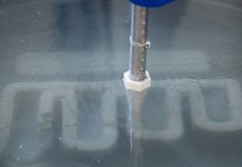
According to researchers, cells employ strategically positioned proteins and mechanical triggers to navigate through their intricate environment, mimicking developmental processes, similar to how the needle of a record player navigates the vinyl grooves to make music.
The team’s most recent study used nanoscale mechanical and chemical cues to mimic cellular functions during growth.
“Our new method serves as an instruction manual for cells, allowing them to create tissues that are better organised and more closely resemble their natural counterparts,” noted Professor Hala Zreiqat.
He underscored that the study is a significant step towards the ability to manufacture functional tissue and organs.
Dr Newman stated that creating tissues from cells required thorough guidance, much like assembling something from many different parts.
“Without specific instructions, the cells would likely group together unpredictably within the incorrect structures. What we’ve effectively done is create a step-by-step process that guides each building block to exactly where it should go and how it should connect with the others,” Dr Newman explained.
In keeping with this approach, Dr Newman said that their recently published work uses a new 3D printing method to define instructions for cells, guiding them to form more organised and accurate structures.
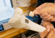
“Through this, we’ve created a bone-fat assembly that resembles the structure of bone and an assembly of tissues that resemble processes during early mammalian development,” he said.
According to the research co-lead, the technology could “revolutionise how we study and understand diseases.”
“By creating accurate models of diseased tissues, we can observe disease progression and treatment responses in a controlled environment. We hope this could one day lead to more effective treatments and even cures for diseases that are currently hard to tackle,” Dr Newman further remarked.
Meanwhile, developmental biologist Professor Patrick Tam who leads the CMRI’s Embryology Research Unit, emphasised that with the new technology, researchers can now control stem cells to become certain cell types and properly organise these cells in time and place, replicating the organ’s real-life development.
The researchers anticipate that the study will pave the way for the treatment of vision loss caused by disorders such as macular degeneration and genetic diseases that cause the loss of retinal photoreceptor cells.
“The idea of treating rare genetic diseases and improving quality of life in this way is empowering. We expect that this work will lead to advanced therapies that can be moved into practice,” Prof Tam stated.
The team said it will now work to improve the process in order to progress the field of regenerative medicine and potentially develop novel treatments for a variety of diseases.


