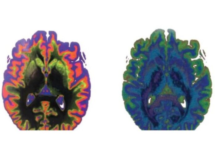
A group of researchers from the University of Colorado has created a new method for converting medical images, such CT or MRI scans, into detailed 3D models on the computer.
In a paper published in the journal 3D Printing and Additive Manufacturing, the scientists describe a method using scan data to create maps of organs made up of billions of volumetric pixels, or “voxels”—like the pixels that make up a digital photograph, only three-dimensional.
The researchers said they are exploring how they can use 3D printers to convert those maps into actual physical models that are more accurate than those that are accessible through existing tools.
According to news released by the university, the discovery is the result of a partnership between researchers at the University of Colorado Boulder and the University of Colorado Anschutz Medical Campus to fill a critical gap in the medical community: Surgeons have long used imaging tools to plan out their procedures before stepping into the operating room.
Robert MacCurdy, assistant professor in the Paul M. Rady Department of Mechanical Engineering at CU Boulder, said his team wanted to fix the gap by giving doctors a new approach to print “realistic and graspable, models of their patients’ various body parts, down to the detail of their tiny blood vessels” using soft and pliable polymers.
MacCurdy, who is also the senior author of the new paper, explained the approach begins with a Digital Imaging and Communications in Medicine (DICOM) file, the standard 3D data that CT and MRI scans produce.
MacCurdy said they turn that data into voxels using specialized software, thereby chopping an organ into tiny cubes with a volume far smaller than a common teardrop.
The team said they built a map for each of the human heart, kidney, and brain structures in order to evaluate these techniques.
The generated maps were sufficiently precise to, for instance, distinguish between the kidney’s fleshy interior, or medulla, and its outer layer or, cortex—both of which appear pink to the human eye.
“Our method addresses the critical need to provide surgeons and patients with a greater understanding of patient-specific anatomy before the surgery ever takes place,” noted MacCurdy.
The project, which is being co-led by MacCurdy and Nicholas Jacobson of CU Anschutz, is being sponsored through AB Nexus, a grant program designed to encourage new collaborations between the two Colorado campuses.


